DENTAL RADIOLOGY
DENTAL RADIOLOGY
In dentistry, X-rays are basically divided into intraoral (the X-ray film is taken inside the mouth) and extraoral (the X-ray film is taken outside the mouth).
Intraoral X-rays are the most commonly used radiological radiographs. These small radiographs provide the dentist with detailed information for caries detection. It is used to obtain information about the tooth and the surrounding bone and assess the tooth’s stage of development.
Extraoral X-rays also show the teeth, but their main focus is on the jawbone and skull. While they do not provide a very detailed view of the teeth, they allow more teeth to be seen at once than intraoral X-rays. With the help of these X-rays, impacted teeth, the developmental status of the jaws, and the temporomandibular joint can be checked.
Panoramic X-Ray
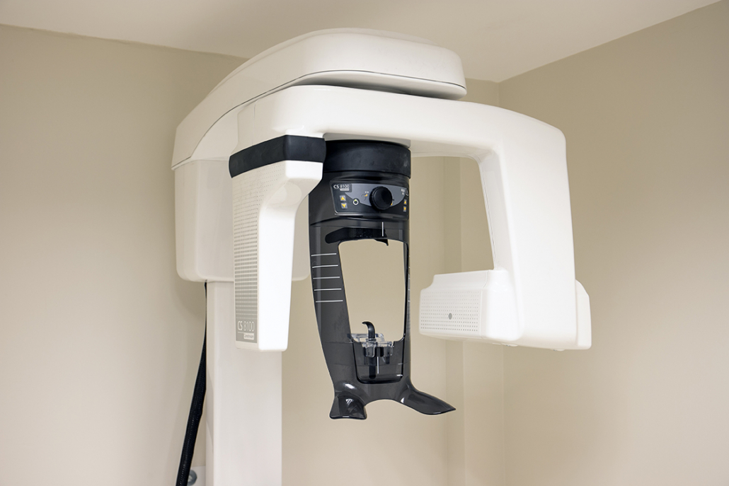
The panoramic X-ray shows the entire oral region. Dentists prefer the evaluation of panoramic X-rays during the initial examination. It is useful in detecting cysts, tumors, impacted teeth, caries. It is a two-dimensional X-ray. Dental tomography, which is recognized in panoramic x-rays and provides three-dimensional and detailed images, is preferred when a more detailed assessment is required.
Dental tomography can be used to assess in detail the bone density, width, and distances to the anatomical points to be considered, such as the mandibular nerve, in the area of the planned dental implant.
Nowadays, dental X-rays are largely used digitally. Thanks to evolving technology, this allows for both faster results and a more detailed image with a smaller amount of radiation.


